The knee meniscus is fibrocartilage that separates the femur from the tibia. We commonly refer it to as cartilage. The knee meniscus has a wedged kidney shape. Each knee joint possesses a medial meniscus and a lateral meniscus. The medial meniscus is an important shock absorber on the medial aspect of the knee joint. It absorbs nearly 50% of the shock of the medial compartment. Thus, when there is a medial knee injury such as a medial meniscus tear, it is very essential to repair the tear, because if not reconstructed and is trimmed out there will be an increase in the load on the medial compartment which finally starts to osteoarthritis and induces medial knee pain.
A medial meniscus tear is more common than a lateral meniscus tear because it firmly attaches to the deep medial collateral ligament and the joint capsule. Also, the medial meniscus absorbs up to 50% of the medial compartment’s shock, making the medial meniscus susceptible to injury.
Meniscus reinforces the rotational stability created by the anterior cruciate ligament. The meniscus likewise acts as a shock absorber. As we walk, run, jump and play sports the knee gets tremendous forces. The meniscus serves to absorb these forces so that the bone surfaces will not damage.
Medial Meniscus Anatomy
The medial meniscus is a fibrocartilage semicircular band that covers the knee joint medially, placed between the medial condyle of the femur and the tibia.
- C-shaped with a triangular cross-section
- Average width of 9 to 10 mm
- The average thickness of 3 to 5 mm
- Composed of fibroelastic cartilage (collagen, proteoglycan, glycoproteins, cellular elements and composed of 65-75% water), collagen (90% Type I collagen), and Fibers (radial and longitudinal).
- The middle genicular artery supply to posterior horns and medial inferior genicular artery supplies peripheral 20-30% of the medial meniscus.
The medial meniscus has a white and red zone. The red zone points out to the outer third of the meniscus. The term red is used to stand for that this zone gets an adequate volume of blood supply. Therefore, meniscus tears in the red zone can frequently heal without surgery. The white zone points out to the inner two-thirds of the meniscus.
Medial Meniscus Tear Causes
A medial meniscus tear can take place from several factors. First, in the younger population, a sporting injury can cause it. Medial meniscus tears commonly happen with an ACL tear by twisting on a slightly flexed knee. This is because the medial meniscus acts as a secondary stabilizer to restrict the knee from slipping forward, and when the ACL tear, it gives extra stress on the medial meniscus which prompts to it tearing. Also, deep squats put extra stress on the back of the knee and can lead to a medial meniscus tear. Other causes include twisting, turning, or pivoting type activities where extra stress put on the medial side of the knee, whereby a medial meniscus tear can take place.
In the older adult, the tear may be because of natural age-related degeneration of the meniscus or a rough arthritic femoral bone surface tearing into the softer meniscus. Here, surgery may be required to attend both the meniscal repair and to repair the damaged joint surface. Depending on the meniscus tear, it may complicate meniscus repair. A large meniscus tear that is inadequately treated may cause degenerative bony (arthritis) changes.
Types of Meniscus Tear
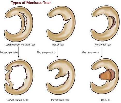
The meniscus can tear anterior to posterior, radially, or can have a bucket handle appearance. A variety of factors are used to determine the ideal treatment of a meniscus tear. Some of these factors include the age of the patient, results of nonsurgical treatment, and if there is another damage than just a torn meniscus.

Also, the specific meniscus tear can determine the most appropriate treatment. Types of meniscus tears-
- Intrasubstance/Incomplete Tear
- Radial Tear
- Horizontal Tear
- Flap Tear
- Complex Tear
- Bucket-Handle Tear
Common tears include bucket handle, flap, and radial.
Medial Meniscus Tear Symptoms
In sports, a meniscus tear typically happens suddenly. Severe pain and swelling may occur up to 24 hours later on. You will additionally experience clicking, popping, or locking of the knee. Walking will become tough. Further pain is also felt when flexing or twisting the knee. A loose piece of cartilage will get stuck within the joints, causing the knee to lock temporarily, restricting the full extension of the leg.
The pain is typically set on the joint’s inner portion, at the joint line, or within the back of the knee, particularly when one squats down. Tears within the front part of the meniscus are less rare; however, these will cause problems in hills or downstairs. Longstanding meniscus tears, or chronic tears, may or may not cause symptoms. Usually, once they cause symptoms, individuals can notice them when they have an extra tear of an underlying tear. There’ll be pain on the joint line with squatting, twisting, turning, or kicking-type maneuvers. Symptoms of medial meniscus tear comprise:
- Pain
- Swelling and stiffness or tightness will increase gradually over 2 to 3 days
- Catching or locking
- Instability
A complex tear of the medial meniscus includes a combination of any of the patterns listed on top of. In several circumstances in patients with these tears, the meniscus has to be trimmed out. However, this will increase the risk of degenerative arthritis, particularly in patients who still take part in impact activities.
How to Diagnose a Meniscus Tear
Your doctor will discuss your symptoms and how the injury occurred. After discussing, your doctor can check for tenderness on the joint line where the meniscus sits. This typically signals a tear.
Medial Meniscus Tear Test
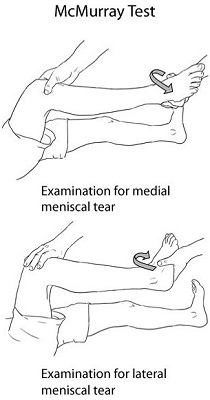
One of the main test for medial meniscus tears is the McMurray test for the knee. Your doctor can bend your knee, then straighten and rotate it. This puts tension on a torn meniscus. If you’ve got a meniscus tear, this movement can cause a clicking sound. Your knee will click each time your doctor does the test.
From a position of maximal flexion, extend the knee with internal rotation (IR) of the tibia and a varus stress.
Internal Rotation (IR) of the tibia + varus stress = lateral meniscus
From a position of maximal flexion, extend the knee with external rotation (ER) of the tibia and valgus stress.

External Rotation (ER) of the tibia + valgus stress = medial meniscus
The IR of the tibia, followed by extension, will check the entire posterior horn to the middle section of the meniscus. The anterior portion of the meniscus isn’t easily tested because the pressure to it part of the meniscus isn’t as great.
Positive findings of the McMurray test meniscus: Pain, snapping, audible clicking or locking will indicate a compromised meniscus.
It is necessary to explain your symptoms accurately. The amount of pain and the first appearance of swelling will offer necessary clues about where and how bad the injury is. Tell your doctor of any recurrent swelling or your knee repeatedly giving way.
Imaging tests
Some other knee issues cause similar symptoms; your doctor may order imaging tests to assist ensure the diagnosis. Although x-rays don’t show meniscus tears, they will show other causes of knee pain, like osteoarthritis.
A magnetic resonance imaging (MRI) scan is commonly used to diagnose menisci injuries. The medial meniscus tear on MRI shows up as black. An MRI is 70 to 90% correct in identifying whether the meniscus has been torn and how badly. However, meniscus tears don’t always seem on MRIs.
It classifies meniscus tears shown by MRI in 3 grades. It does not consider grades 1 and 2 are serious. They may not even be clear with an MRI examination. Grade 3 is a true meniscus tear, and an arthroscope is close to 100 percent correct in diagnosing this tear.
Recommended Post
- UFlex Athletics Knee Compression Sleeve Reviews
- Top Rated Mueller Knee Brace for Knee Support
- Top-Rated DONJOY ACL Knee Braces
- The 5 Best Hinged Knee Brace Reviews
Treatment for a Medial Meniscus Tear
Meniscus tear healing depends on your meniscal blood supply, which is limited. Meniscus gets its nutrition from blood and synovial fluid within the joint capsule. Your meniscus has two distinct regions that affect their ability to heal. We call these the Red Zone and the White Zone.
Red Zone
The red zone encompasses a blood supply, which implies that they will heal naturally. A “longitudinal tear” is an example of a meniscal tear that is probably going to heal naturally. The outer edge of the meniscus has the blood supply from the synovial capsule. Lateral meniscal tears may heal with no surgery.
White Zone
The white zone doesn’t have a blood supply and won’t heal naturally. This meniscus receives its nutrition from the synovial fluid. Because of this, tears of the inner meniscus rarely heal because of an absence of blood supply to trigger an inflammatory response. These injuries usually need surgery to remove the torn part.
Nonsurgical Treatment
If your MRI shows a Grade 1 or 2 tears, however, your symptoms and physical exam are inconsistent with a tear; it may not need surgery. As long as your symptoms don’t persist and your knee is stable, nonsurgical treatment may be all you need.
The RICE protocol is effective for many sports-related injuries. RICE stands for Rest, Ice, Compression, and Elevation.
- Take time off from the activity that led to the injury. Your doctor may suggest that you use crutches to avoid putting weight on your leg.
- Use cold packs for 20 minutes at a time, many times daily. Don’t apply ice directly to the skin.
- To prevent further swelling and blood loss, wear an elastic bandage (compression).
- To reduce swelling, recline once you rest, and place your leg up on top of your heart.
Non-steroidal anti-inflammatory medicines like aspirin and ibuprofen cut back pain and swelling. A small meniscus tear, or a tear in the red zone, can sometimes respond quickly to physiotherapy treatment. One of the main roles of your meniscus is shock-absorption. Luckily, the alternative vital shock absorbers around your knee are quads and hamstring muscles.
Researchers have recognized that if you strengthen your quads and hamstring muscles, your bone stresses can diminish as your muscle strength improves and your knee becomes more dynamically stable.
Medial meniscus tear physical therapy treatment can aim to:
- Reduce pain and inflammation.
- Normalize joint range of motion.
- Strengthen your quad and hamstrings.
- Strengthen your calves, hip, and pelvic muscles.
- Normalize your muscle lengths that will improve your proprioception, balance, and function eg walking, running, squatting, hopping and landing.
- Improve patellofemoral alignment.
- Minimize your probability of re-injury.
Meniscal injuries are usually related to other knee injuries like ACL, which need to be treated with your meniscal tear.
Medial Meniscus Tear Brace
Wearing a rehabilitative knee brace will help recover from a torn meniscus by adding support to the joint and limiting the wearer from provoking the injury even further. Knee braces with hinges on each side of the knee offer stability and prevent that wobbly feeling you may get after a knee injury. Medial unloader knee braces provide excellent support in this condition.
Meniscus Tear Exercises
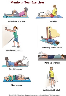
Physical therapy and home exercises can advance healing in your knee and help you return to desired activities. Develop strength and flexibility in your knee and legs may further prevent future degeneration in your knee. The following exercises for meniscal tears assist you to strengthen your lower limb muscles-
- Quad sets
- Hamstring curls
- Straight-leg raises to the back
- Straight-leg raises to the front

- Heel raises
- Heel dig bridging
- Standing knee bends
Meniscus Tear Exercises to Avoid
Avoiding twisting movements for a torn meniscus. One should perform quadriceps setting exercises with the knee straight or mini-squats, bending the knee only 15 degrees, to prevent quadriceps muscle atrophy.
How Long for a Meniscus to Heal Without Surgery?
Your meniscal tear can usually take up to six or eight weeks to completely heal if the injury is within the red zone. As mentioned previously, some meniscal tears would require surgery. Your physical therapist can guide you on what’s most likely for your knee injury.
It is necessary to avoid activities and exercises that place excessive stress through your meniscus and further delay healing. Sometimes, your physical therapist may advise you to stay weight off your knee. In these instances, it’s going to recommend crutches. Everyone seems to be different, therefore, be guided by your physical therapist.
Surgical Treatment
Grade 3 meniscus tears usually need surgery, which can include:
- Arthroscopic Repair — an arthroscope is inserted into the knee to visualize the tear. It makes one or two for inserting instruments. It repairs any tears with dartlike devices that are inserted and placed across the tear to carry them together. Arthroscopic meniscus repairs generally take about 40 minutes. Usually, you may leave the hospital on the same day.
- Arthroscopic Partial Meniscectomy — The goal of this surgery is to get rid of a small piece of the torn meniscus to induce the knee functioning normally.
- Arthroscopic Total Meniscectomy — sometimes, arthroscopic total meniscectomy, a procedure in which it removes the whole meniscus will treat a large tear of the outer meniscus.
The best treatment choice is to repair the torn meniscus and save the maximum shock absorber as possible. This can leave you with close to “normal” structures and reduce the chance of degenerative arthritic changes in later life.
Post-Surgical Physical Therapy for Meniscal Injuries
The posterior horn of the medial meniscus is the posterior third of the medial meniscus. It placed in the back of the knee. It is the thickest portion and absorbs the most force; Therefore, it contributes the most stability to the knee and is the most vital segment of the medial meniscus.
For young active persons with grade 3 tears in the posterior horn of the medial meniscus, the meniscal repair is the best option. Repair of the meniscus gives the best possible chances for an asymptomatic knee in the long run. If the tear is in an older individual such as above 50, and if it is a degenerative tear with no symptoms like locking, can try the conservative treatment.
Resected Meniscal Tears
Physiotherapy rehabilitation for resected meniscal tears will usually be moderately aggressive, targeting early come back to function. It’ll progress you through rehabilitation as your pain and swelling allow. Most arthroscopic patients will return to normal function within 3 to 6 weeks.
Post-Meniscal Repair
Rehabilitation after a meniscus repair is typically different from a resection because of the healing time needed where a meniscus has been stitched. Most surgeons can like non-weight bearing for 4 to 8 weeks to permit the meniscus to heal before starting weight-bearing exercises.
Physiotherapy rehabilitation should concentrate on the early mobilization of the knee (tibiofemoral) and kneecap (patellofemoral) joints, and strengthening of your quad, hamstrings, and leg muscles.
Your treatment guidelines are just like the nonoperative approach, taking into consideration the findings and operative procedures performed. For more specific data, please ask your physical therapist.
Meniscus Tear Surgery Recovery Time Back to Work
The approach for patients who go through a partial medial meniscectomy is to begin physical therapy on the first day after surgery. A treatment procedure acting on the reactivation of the quadriceps muscles, recovering full knee and patellar mobility, and an effective resolution of knee swelling is emphasized. It recommends that patients who have a minimum amount of meniscus trimmed out take back on any impact activities until a minimum of 6 weeks after surgery. In cases that have a notable amount of meniscus resected, it is recommended to avoid significant impact activities because of the greater risk of the development of osteoarthritis in these cases with this activity.
You might return to a physically active job in four to six weeks. For a very physically active role or to come back to sports, arrange on a three- to the six-month recovery period.
What Happens to Untreated Torn Meniscus?
An untreated meniscus tear will deteriorate and may come loose within the knee joint. This will cause knee locking or giving way. Untreated meniscus tears also can increase tear size and extend the damaged area of the meniscus. And increase the chance of degenerative arthritic changes in later life.
